Iron deficiency (ID) is the most common nutritional deficiency worldwide. While most efforts target iron deficiency anemia, ID without anemia is also common and carries important health consequences. Traditional diagnostic methods rely heavily on hematologic markers like hemoglobin or bone marrow iron, often overlooking effects of low iron levels on other organs—especially the brain.
Iron is important for brain development, including neurogenesis, myelination, and neurotransmitter production. Even without anemia, low iron levels have been linked to psychiatric issues, sleep disturbances, and cognitive deficits. Yet, the current diagnostic cutoffs often ignore these neurological effects. Recent advances in imaging techniques, particularly quantitative susceptibility mapping (QSM) in magnetic resonance imaging (MRI), allow indirect measurement of brain iron levels. QSM is especially useful for assessing iron in gray matter areas like the basal ganglia, where iron is abundant and vital for function. This opens new pathways for understanding how subclinical iron deficiency may impact brain health in the youth.
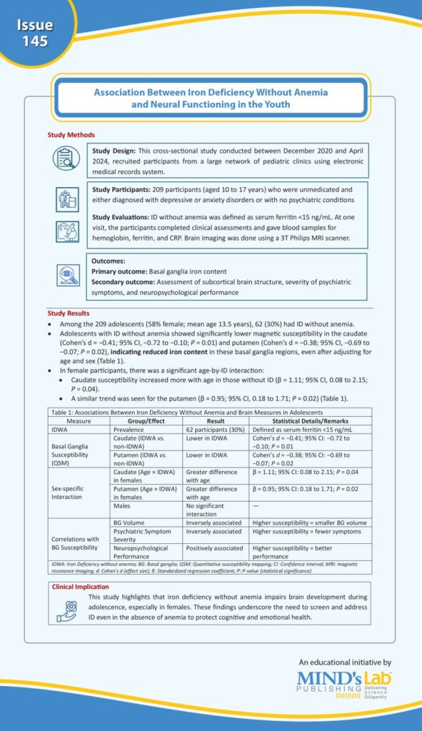
A study by Fiani et al., published in the journal “JAMA Network Open”, aimed to investigate the association between iron deficiency (ID) without anemia and basal ganglia (BG) iron content, its structure, and function in adolescents. In this cross-sectional study, 209 unmedicated participants aged 10–17 years (58% female) were enrolled between 2020 and 2024. ID without anemia was defined as serum ferritin <15 ng/mL. MRI was used to assess BG susceptibility and volume, along with psychiatric interviews and cognitive testing. ID without anemia was observed in 62 participants (30%). This group had significantly lower susceptibility in the caudate (d = −0.41; P = 0.01) and putamen (d = −0.38; P = 0.02). Among female subjects, increasing age amplified this difference. Lower BG iron levels were linked to higher psychiatric symptoms and poorer neurocognitive performance. This study outlines the need to consider brain health in the clinical assessment of ID as ID without anemia may negatively impact brain development and function during adolescence (a period of brain development), especially in females (See Graphic).

(Source: Fiani D, Kim JW, Hu M, Salas R, Heilbronner S, Powers J, Haque M, Dinh S, Huang X, Worthy D, Devaraj S. Iron deficiency without anemia and reduced basal ganglia iron content in youths. JAMA Network Open. 2025;8(6):e2516687- Doi:10.1001/jamanetworkopen.2025.16687)
